|
|
Appendix 1 - Somatic Sensory pathways |
||
| Suggested readings from Neuroscience, 5th ed. |
|
|
Appendix 1 Overview
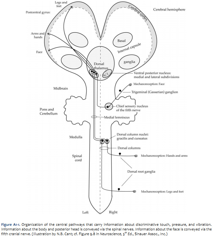
IntroductionThe goal of this Appendix is to review the functional organization of the somatic sensory system, from the activation of the peripheral receptors to the discharge of neurons in the somatosensory cortex. Many of the structures involved in these processes have been identified in Labs 3 & 5. Here, our aim is to bring together the relevant neuroanatomy for understanding somatic sensation, and for localizing components of the pathways involved in sectional views of the CNS. A more detailed account of the physiology of these processes is provided in Purves et al., Neuroscience 5th Ed., Chapters 9 & 10 (Sinauer Assoc., Inc.). There are two major, parallel systems that convey somatosensory information from the periphery of the post-cranial body to the cortex, the dorsal column-medial lemniscus system and the anterolateral system. There are comparable parallel systems carrying information from the face associated with the central projections of the trigeminal nerve. In addition, there is an important system carrying proprioceptive information from the muscle spindles to the cerebellum and cortex. The following sections review the pathways taken by the components of each of these systems. It is important for your understanding of neurological deficits seen in the clinic to know where these pathways travel relative to each other and to other structures (including the cranial nerve nuclei) in the brain. Pathways mediating mechanoreceptionThe pathways that convey information about touch (especially fine, discriminative touch), pressure, and vibration from the body and face are illustrated in Figures A1-1 and A1-2. In the dorsal column-medial lemniscal system, which carries information from the body, the first order neuron is the dorsal root ganglion cell. The first-order cells have peripheral processes that are often encapsulated (e.g., Meissner corpuscles, Pacinian corpuscles) or are associated with specialized receptor cells (e.g., Merkel’s discs). They send a central process into the spinal cord, where a major branch travels in the dorsal columns to the caudal end of the medulla. There, it synapses on neurons in the dorsal column nuclei, the gracile and cuneate nuclei. The second-order neurons located in the dorsal column nuclei send their axons across the midline, where they form a fiber bundle known as the medial lemniscus (hence, the name of this pathway). The axons of the medial lemniscus run through the rest of the medulla, the pons and the midbrain before ending in the ventral posterior lateral nucleus (VPL) of the thalamus. Third-order neurons in the VPL send their axons via the internal capsule to terminate in the somatic ssensory cortex, which lies in the postcentral gyrus. The first-order neurons associated with receptors in the face lie in the trigeminal (Gasserian) ganglion. The central processes enter the brain via the fifth cranial nerve and terminate in the principal (or chief) sensory nucleus of the trigeminal complex (or of the fifth nerve). The second-order cells in this nucleus send axons across the midline to form the dorsal trigeminothalamic tract, also known as the trigeminal lemniscus. These second-order axons travel to the ventral posterior medial nucleus (VPM) of the dorsal thalamus. Third-order neurons in the VPM send their axons to the postcentral gyrus. Pathways mediating pain and temperature sensation.
The anterolateral system is responsible for conveying information about pain, temperature and crude touch (i.e., touch lacking the spatial resolution of the dorsal column system) from the post-cranial body. Comparable information about the face is processed in trigeminal pathways. These pathways are illustrated in Figures A1-3 and A1-4. Most peripheral processes associated with the dorsal root ganglion cells that contribute to this system are “free.” That is, they are not associated with encapsulated endings like those in the dorsal column-medial lemniscal system. In addition, the first-order fibers associated with the anterolateral system are generally much smaller in diameter than those associated with the dorsal column system. (So what does this tell you about the relative conduction velocities of these two important somatic sensory pathways?) The first-order neurons in the anterolateral system, like those in the dorsal column-medial lemniscal system, have their cell bodies in the dorsal root ganglia. The central processes of these neurons terminate on second-order neurons in the dorsal horn of the spinal cord. Pain and temperature information from receptors in the face is carried into the brain on the fifth nerve. The cell bodies of the first order neurons are in the trigeminal ganglion and the central processes of the cells make synapses in a nucleus in the medulla known as the spinal trigeminal nucleus (of the fifth nerve). This nucleus is actually continuous with the dorsal horn of the spinal cord (this explains the “?” in Figure 5.8). The second-order neurons in the dorsal horn of the spinal cord send their axons across the midline, where they accumulate in the anterolateral (ventrolateral) part of the white matter. They ascend in this location through the length of the cord. Many of these fibers continue through the medulla, the pons and the midbrain to contact third-order neurons in the ventral posterior lateral (VPL) nucleus of the thalamus (as well as other thalamic nuclei). This direct pathway from the spinal cord to the thalamus is often called the spinothalamic tract. Actually, the thalamus is only one of the targets of the second-order neurons in the anterolateral system. These neurons also project to central parts of the medulla, pons and midbrain (known collectively as the reticular formation) and to the periaqueductal gray matter and the superior colliculus. (These pathways are not illustrated on Figures A1-3 or A1-4.) Second-order neurons located in the spinal trigeminal nucleus send their axons across the midline to form the ventral trigeminothalamic tract, which travels to the ventral posterior medial (VPM) nucleus of the thalamus. Third-order neurons in the ventral posterior nucleus and in other thalamic nuclei then project to the cortex via the internal capsule. The postcentral gyrus appears to be important for the ability to discriminate the exact location of painful stimuli, but many other, less well-understood cortical areas (including areas in the anterior part of the cingulate gyrus) appear to be important in the complete sensation of pain, including the complex affective dimensions of pain. Figure A1-5 presents a diagram of the major parallel pathways carrying somatic sensory information to the cerebral cortex. The pathways for mechanoreception shown in Figure A1-1 and the pathways for pain and temperature sensation shown in Figures A1-3 are shown together bilaterally.
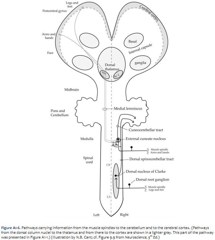
Pathways mediating proprioception
Cutaneous receptors that feed into the dorsal column-medial lemniscal system are one source of information giving us a sense of body position. A second important source of such information is the pathway leading from muscle spindles. Two important targets of this information are the postcentral gyrus and the cerebellar cortex, as illustrated in Figure A1-6. The afferent fibers associated with muscle spindles in the legs send their central processes into the spinal cord where they form synapses in a prominent nucleus in the thoracic and upper lumbar spinal cord, known as the dorsal nucleus of Clarke. (The axons ascend in the dorsal columns from their point of entry until they reach the thoracic cord, where they form synapses in this nucleus.) The second-order neurons in Clarke’s nucleus send very large, heavily myelinated axons into the dorsolateral white matter on the same side, where they ascend as the dorsal spinocerebellar tract. A major target of the spinocerebellar tract, as its name implies, is the cerebellum. The fibers enter the cerebellum via the inferior cerebellar peduncle. Branches of the spinocerebellar tract also form synapses on cells in or near the dorsal column nuclei, which appear to then send axons into the medial lemniscus (joining the pathway already illustrated in Figure A1-1). The afferent fibers associated with muscle spindles in the arms send their central processes into a nucleus at the caudal end of the medulla, known as the external cuneate nucleus (because it lies just lateral to the cuneate nucleus, already described). The second-order cells send axons into the cerebellum on the same side via the cuneocerebellar tract, which—like the dorsal spinocerebellar tract—enters the cerebellum via the inferior cerebellar peduncle. The functions of these pathways are:
The locations of major components of these pathways are indicated in Figure A1-7.
|
||
Click here to submit questions or comments about this site. Updated 1/10/11 - Velkey |
||